
Showing posts with label microscopy. Show all posts
Showing posts with label microscopy. Show all posts
Thursday, February 9, 2012
Thursday, February 2, 2012
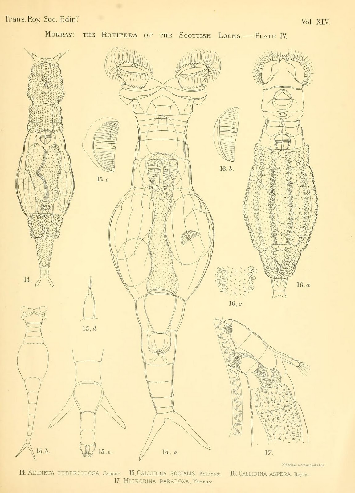
"The Rotifera of the Scottish Lochs"
Thursday, January 26, 2012
Sand, magnified

Monday, January 9, 2012
Planktonic protists
The word "plankton" shares a Greek root with "planet" (πλαγκτός,
meaning drifting or wandering)-- an etymological link which seems
particularly appropriate for these drifting microbes, spherical or
stellate in form:
This beautiful clip is one of a series called Plankton Chronicles, found via The Book of Barely Imagined Beings.
This beautiful clip is one of a series called Plankton Chronicles, found via The Book of Barely Imagined Beings.
Friday, January 6, 2012
Sponges and their skeletons
Images of sea sponges from the Report on the Hexactinellida collected by H.M.S. Challenger during the Years1873-76; each image shows a sponge framed by examples of the glass-like spines (called spicules) which make up its "skeleton":


Sea cucumbers also have attractive mineralized parts called spicules, but these function as external armor rather than internal support. 

Wednesday, January 4, 2012


Wednesday, December 21, 2011
Characodictyon

Image from Live Science.
Saturday, December 17, 2011
Micro-plants
Illustrations of algae, plants, seaweed, and fungi from Common Objects of the Microscope, by John George Wood, 1900:


Saturday, December 10, 2011
Diatom mandala
This beautiful design fits on a microscope slide, and the jewel-like objects that compose it are the shells of tiny marine microbes.

Image from Klaus Kemp's diatom art site. Diatom arrangements like these, along with similar arrangements of butterfly scales, were popular in the Victorian era.
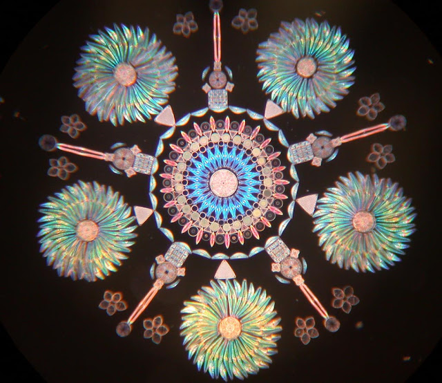
Image from Klaus Kemp's diatom art site. Diatom arrangements like these, along with similar arrangements of butterfly scales, were popular in the Victorian era.
Saturday, December 3, 2011
Fairy wasp

[above] The fairy wasp, Megaphragma mymaripenne... pictured next to a Paramecium and an amoeba at the same scale.From Not Exactly Rocket Science.
...
As they get smaller, insects can do away with many of their organs. The feather-winged beetles – twice as big as the fairy wasps, but still impressively tiny – have drastically reduced the size of their genitals, guts and breathing tubes. They have totally lost their hearts: at their size, diffusion is enough to carry liquids around their body without the need for a pump. Their wings, like those of thrips and fairy wasps, are little more than wispy strands, rather than the flat oars of most other insects. That’s all they need to paddle through thick air currents.
Monday, November 7, 2011
Infusorial Animalcules

Plate from Pritchard's 'Infusorial Animalcules' 1852 showing illustrations of freshwater microscopic organisms.
Wednesday, November 2, 2011
Starfish larva

Even stranger is the starfish Luidia-- its larva splits into a juvenile starfish and a gelatinous planktonic creature, which go on to live independent lives.
Monday, October 24, 2011
Butterfly egg

David Millard,Vanessa atalanta (Red admiral butterfly) egg in Urtica dioica (Stinging nettle) trichomes (10X)From Nikon Small World.
Thursday, September 29, 2011
Pediastrum
A beautiful pond-dwelling microscopic algae:
 Image from MicroscopyUK, which has many more photos of similar algae.
Monday, September 19, 2011
Oolite

Monday, September 5, 2011
"Feathers of Humming Bird, Brittle Star Fish, Fossil Tooth of Shark"

These slides have cover slips of thin glass, which was very expensive and difficult to produce before the 1840s—early mounters more often used sheets of mica, which was far from transparent. The use of Canada balsam sap (which preserves structures and eliminates air and water from samples) as a mounting medium also vastly improved the view.From a SEED Magazine slideshow of Victorian microscope slides. Those shown above are from the collection of Howard Lynk, who has many more; the arranged slides of diatoms and spicules are quite lovely.
Monday, July 11, 2011
Foraminifera

Sunday, June 19, 2011


Thursday, May 26, 2011

Wednesday, March 9, 2011
Spicules

Illustrations of spicules, spines found in the skin of sea cucumbers.
Micro-photos of these pretty objects can be seen here and here; they also appear in the border of this sea-cucumber drawing by Ernst Haeckel. 
Subscribe to:
Posts (Atom)